…
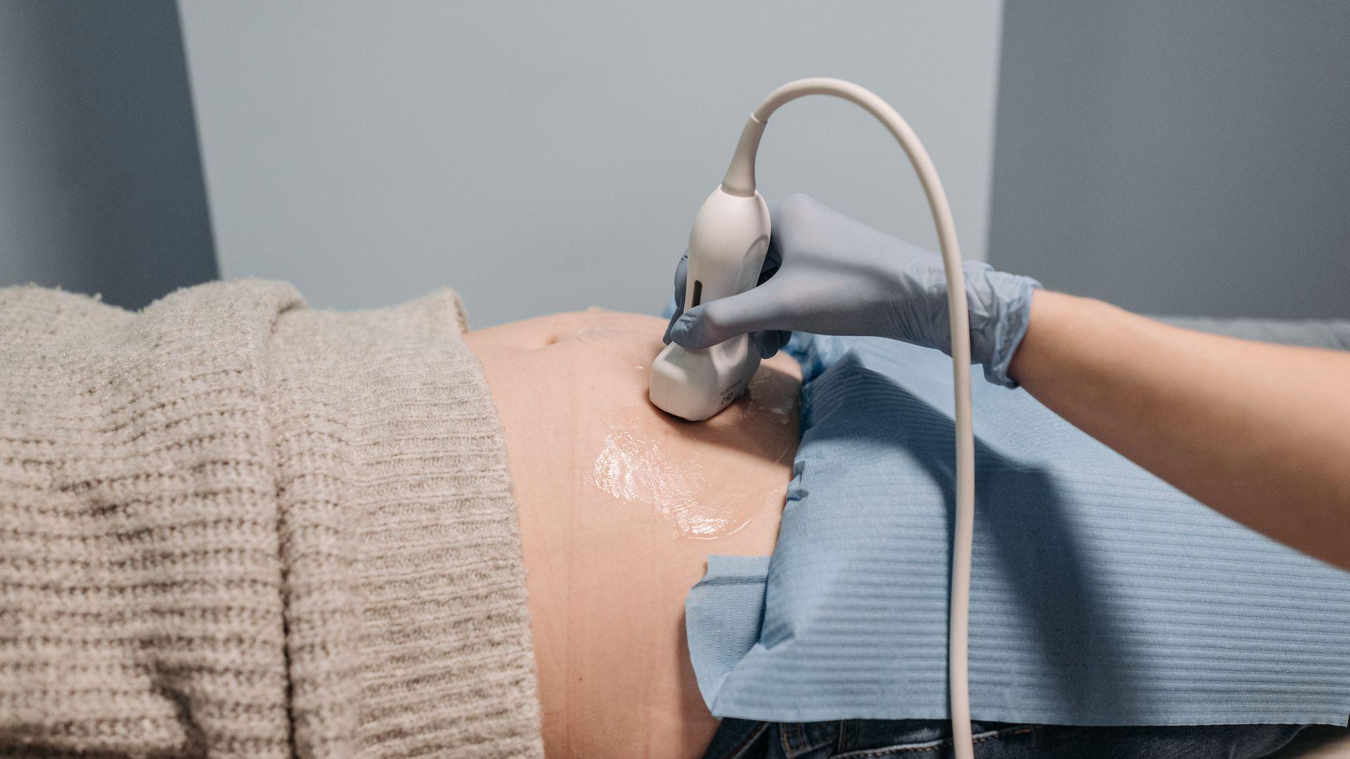
Prenatal screening
Prenatal screening is a series of tests during pregnancy that concern both the baby and the mother. Its purpose is a smooth course of pregnancy and the early detection of problems.
Who it is for
In the past, prenatal screening was thought necessary only in a few special cases. Today it is accepted that every pregnant woman should have it, regardless of age, family history, number of pregnancies or origin.
The tests fall into two groups: ultrasound and invasive. Ultrasound screening applies to all pregnant women. Invasive testing is suggested only when more specific investigation is needed.
What we look for
Prenatal screening detects abnormalities in the baby's development and medical conditions in the mother, at specific stages of pregnancy. Abnormalities in the baby may be structural, that is, affecting the formation of organs such as the skull, brain, face, spine, limbs, chest and abdomen. These are diagnosed directly on ultrasound.
They may also be chromosomal, such as Down syndrome (trisomy 21), Edwards syndrome (trisomy 18), trisomy 13 or Turner syndrome. These are detected indirectly, by observing certain features of the baby that we call markers. In the mother, we check for conditions that may cause complications, such as high blood pressure, the risk of preterm birth and reduced growth of the baby.
Ultrasound screening by trimester
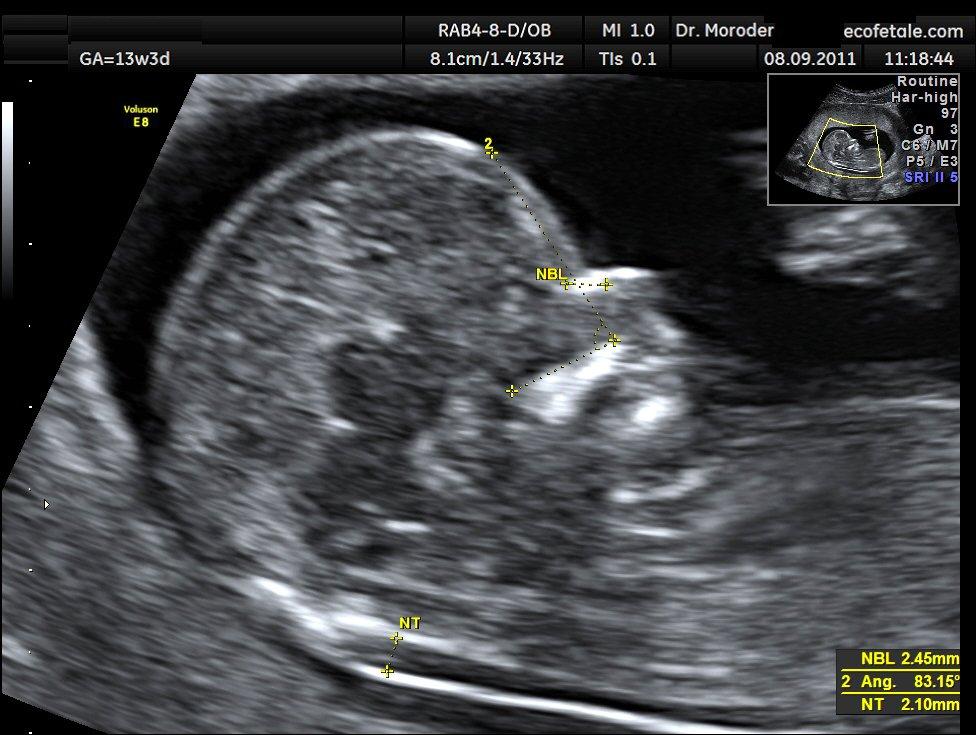
In the first trimester (weeks 6–14) we confirm that there is a pregnancy and where it is, inside or outside the womb, the number of babies, the heartbeat and any serious structural abnormalities. Between 11 and 14 weeks the nuchal translucency scan is performed, which has its own page.
In the second trimester, between 20 and 24 weeks, the detailed anomaly scan (level II scan) is performed. It includes a thorough check of the baby's anatomy and of the markers for chromosomal abnormalities. Where there is an indication, such as a history of heart disease or diabetes, a scan of the baby's heart is recommended between 22 and 24 weeks. Colour Doppler is used to check blood flow in the uterine arteries, a predictive sign for high blood pressure in pregnancy. A transvaginal scan measures the length of the cervix and, with a vaginal swab culture, we assess the risk of preterm birth.
In the third trimester, after the 28th week, the biophysical profile is performed to confirm the baby's normal growth and wellbeing. We examine the position and appearance of the placenta, the amount of amniotic fluid, the head and abdominal circumferences and the thigh bone, from which the estimated weight is calculated. We also observe the baby's movements, breathing movements and muscle tone, and record its heartbeat with a CTG. In high-risk pregnancies we use Doppler to measure blood flow in the uterine, umbilical and cerebral arteries, for the early detection of placental insufficiency.
Invasive tests
When the scan shows positive markers for chromosomal abnormalities, when the mother is over 35, or when there is a significant family history of chromosomal abnormalities, blood disorders such as thalassaemia, or metabolic diseases such as cystic fibrosis, the investigation is extended to invasive methods. These give a diagnostic answer with very high accuracy, but carry a small risk of miscarriage, about 1%.
Chorionic villus sampling (CVS) is done between 11 and 14 weeks. A small tissue sample is taken from the placenta for a karyotype, that is, a chromosomal analysis of the baby, with the result in about 3 days. Amniocentesis is done after the 15th week, using a sample of amniotic fluid, and the result takes about 15 days. Less often, fetal blood is taken from the umbilical cord.
The rest of this page is available in Greek. View the Greek page
Frequently asked questions
I am young and have no family history. Do I need prenatal screening?
Yes. Ultrasound screening applies to every pregnant woman, regardless of age, history or number of pregnancies. Invasive tests, on the other hand, are suggested only when there is a specific reason. We will explain the schedule of scans at your first visit.
Do I need to drink water before the scan?
No. Ultrasound screening in pregnancy does not require you to drink fluids beforehand and is not invasive. Simply come as you are, with the results of your previous tests.
When is amniocentesis suggested?
When the scan shows positive markers for chromosomal abnormalities, when the mother is over 35, or when there is a family history of chromosomal abnormalities, thalassaemia or cystic fibrosis. It is done after the 15th week and gives the baby's karyotype in about 15 days. It carries a small risk of miscarriage, about 1%, which is why the decision is made together with you, after weighing the facts.
What is the level II scan?
It is the detailed scan of the second trimester, between 20 and 24 weeks. It checks the baby's anatomy thoroughly, along with the markers for chromosomal abnormalities. In the same period, where indicated, a scan of the baby's heart is also done. We will tell you in good time when to schedule it.